Janelia Fluor® Dyes
Originally developed at the Janelia Research Campus, the Janelia Fluor® dyes have recently been made commercially available conjugated to antibodies or supplied as NHS esters for custom labeling. These bright, photostable dyes enable sensitive detection of cellular targets in IHC, ICC, and flow cytometry applications. They are ideal for Super-Resolution Microscopy (SRM) and are especially suited for live-cell imaging.
Janelia Fluor® Products
Janelia Fluor® Dyes
Janelia Fluor® Dyes
- Exceptionally bright, highly photostable
- Especially well-suited to live-cell imaging
- Ideal for Super Resolution Microscopy (SRM) including dSTORM and STED
- Suitable for use in confocal microscopy, IHC, ICC and flow cytometry
- Spectra Viewer tool available for multiplex experimental design
- Spontaneously Blinking Janelia Fluor® dyes, ideal for single-molecule localization microscopy (SMLM)
- Photoactivatable Janelia Fluor® dyes also available, compatible with photoactivated localization microscopy (PALM)
Janelia Fluor® Conjugated Antibodies
Janelia Fluor® Conjugated Antibodies
- We offer Janelia Fluor® conjugated primary and secondary antibodies: JF 549 Antibodies and JF 646 Antibodies
- Antibody custom conjugation service also available
Janelia Fluor® Based Cellular Stains
Janelia Fluor® Based Cellular Stains
Fluorogenic, enabling no-wash experiments, with bright, low noise signals, perfect for sensitive or low expression experiments.
We offer Janelia Fluor® based probes for staining cellular structures and organelles, including:
- Taxol Janelia Fluor® 646 for direct imaging of the microtubule cytoskeleton
- Hoechst Janelia Fluor® 646, a red-emitting DNA probe
- Pepstatin A Janelia Fluor® 526, to track and stain lysosomes
Janelia Fluor® dyes are available with a variety of reactive handles for biomolecule conjugation. Conjugation protocols are available as well as Custom Chemistry services for alternative reactive handles.
Janelia Fluor is a registered trademark of Howard Hughes Medical Institute.

- "We have been using the JF secondary antibodies and like them a lot for IHC. As advertised, they seem to be at least a little brighter and more photostable than the Alexa 647 and Cy3 dyes we had been using. We will probably switch over to these completely going forward." - Steve Stowers, PhD, Assistant Professor, Montana State University
- "It’s a great antibody and great fluorophore by its signal sharpness and photo stability." - Anonymous
Human tonsil stained for aSMA (red; alpha-Smooth Muscle Actin Antibody [Janelia Fluor® 549] Catalog # NBP2-34522JF549) and counterstained with DAPI (blue).
Luke Lavis: Bright Ideas in Fluorescence Imaging
In this episode of the Back of the Napkin podcast, discover how the work of Luke Lavis, at the Janelia Research Campus, has pushed the boundaries of fluorescence imaging. His development of Janelia Fluor® dyes has significantly improved live-cell and super-resolution imaging capabilities.
This podcast reveals the intricate balance between fundamental research, application in drug discovery, and the collaborative culture at Janelia, emphasizing the importance of mentorship and interdisciplinary interactions.
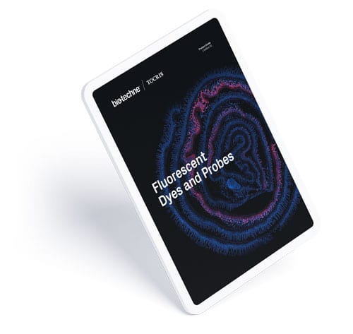
Fluorescent Dyes and Probes Brochure
This product guide showcases our range and gives background on the use of Fluorescent Dyes and Probes.
Immunohistochemistry (IHC) Handbook
This recently updated handbook offers a comprehensive introduction to IHC.
-
Grimm et al. (2015) A general method to improve fluorophores for live-cell and single-molecule microscopy Nat.Methods 244:12. PMID: 25599551.
-
Grimm et al. (2016) Bright photoactivatable fluorophores for single-molecule imaging Nat.Methods 13:985. PMID: 27776112.
-
Grimm et al. (2017) A general method to fine-tune fluorophores for live-cell and in vivo imaging Nat.Methods 14:987. PMID: 28869757.
-
Grimm et al. (2017) General Synthetic Method for Si-Fluoresceins and Si-Rhodamines ACS Cent.Sci. 3:975. PMID: 28979939.
-
Lavis et al. (2017) Chemistry Is Dead. Long Live Chemistry! Biochemistry 56:5165. PMID: 28704030.
-
Tsai et al. (2017) Nuclear microenvironments modulate transcription from low-affinity enhancers Elife 6:e28975. PMID: 29095143.
-
Yadav et al. (2017) Bright Dyes Bring Biology into Focus ACS Cent.Sci. 3:920. PMID: 28979930.
-
Zhang et al. (2017) Optogenetic control of kinetochore function Nat.Chem.Biol. 1096:13. PMID: 28805800.


