The Role of Caspase-3 in Apoptosis
Caspase-3 enzyme is a member of the family of endoproteases which regulate inflammation and apoptosis signaling networks. Caspase-3 is known as an executioner caspase in apoptosis because of its role in coordinating the destruction of cellular structures such as DNA fragmentation or degradation of cytoskeletal proteins (1). The activity of caspase-3 is tightly regulated and it is produced as zymogen in an inactive pro-form (1). Caspase-3 antibodies serve as excellent biomarkers to monitor induction of apoptosis by detecting the levels of pro caspase-3 and its active form. Cleavage and activation of pro caspase-3 is catalyzed by caspase-8, caspase-9, and granzyme B to generate the active heterodimer of caspase-3 subunits (1).

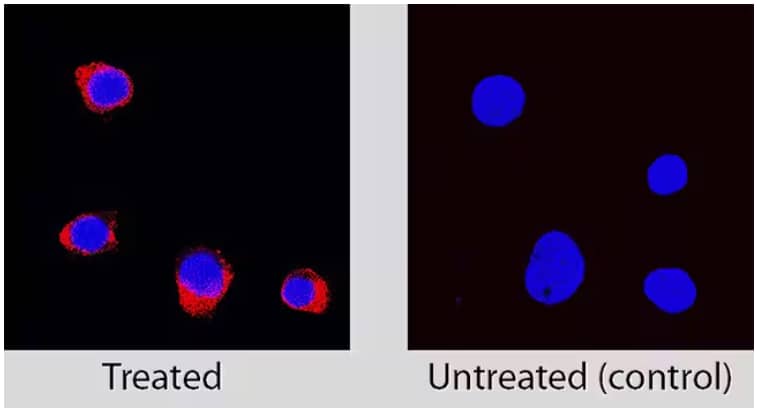
Orthogonal Strategies Validation. Immunocytochemical detection of Caspase‑3 in immersion fixed Jurkat human acute T cell leukemia cell line treated with Staurosporine, an apoptosis inducing agent, using Rabbit Anti-Human Caspase‑3 (1112I) Monoclonal Antibody (Catalog # MAB7071), followed by staining cells with NorthernLights™ 557-conjugated Anti-Rabbit IgG Secondary Antibody (red; Catalog # NL004) and counterstained with DAPI (blue). Specific Caspase-3 antibody staining was localized to cytoplasm.
Apoptosis Assays and Caspase-3 Antibodies
Lin et al. used caspase-3 antibodies to monitor the induction of apoptosis and caspase-3 cleavage and activation through western blotting (2). They showed cells infected with Helicobacter pylori are sensitized to TNF-related apoptosis-inducing ligand (TRAIL)-mediated apoptosis (2). Polymorphisms in the caspase-3 coding region have been shown to lead to increased cancer susceptibility tumorigenesis for multiple myeloma, squamous cell carcinoma, and lung cancer (1). In a study of small cell lung cancer, the experimental drug veliparib and its effect on cells in conjunction with chemotherapy (3). This study used cleavage of caspase-3 as measured through western blotting with the caspase-3 antibody to show veliparib potentiates sensitivity to chemotherapeutic agents (3). Caspase-3 antibodies have proven to be valuable tools in mechanistic investigations of new cancer drugs. Chen et al. investigated the experimental drug moscatilin and used caspase-3 antibodies to examine the ability of moscatilin to inhibit proliferation of colorectal cancer cells and induce apoptosis (4).

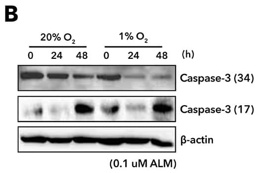
Biological Strategies Validation. Western Blot analysis of PC3 cells treated with 0.1 μM ALM for 24 h and 48 h under normoxia (20% O2) and hypoxia (1% O2) and probed with Mouse anti-Caspase-3 Monoclonal Antibody (CPP32 4-1-18) (Catalog # NB500-210). Results show that ALM induces HIF-1alpha-dependent apoptosis, and an increase in cleaved Caspase-3, in PC3 cells. Image collected and cropped by CiteAb from the following publication (//www.mdpi.com/2073-4409/8/5/439), licensed under a CC-BY license.
Bio-Techne offers Caspase-3 reagents for your research needs including:
- Caspase-3 antibodies
- Caspase-3 antibody pairs
- Caspase-3 ELISA kits
- Caspase-3 kits
- Caspase-3 lysates
- Caspase-3 proteins
- Caspase-3 RNAi
PMIDs
- 23545416
- 24603337
- 25124282
- 18594007
Note: Some headings, titles, links, and images in the blog were updated in January 2023