By Ruha Adelkar, M.S.| MBA
Immunohistochemistry (IHC) is a pivotal technique in the life sciences, enabling researchers and clinicians to visualize protein expression within tissue sections. Traditional methods, however, often come with challenges such as non-specific staining and time-consuming protocols. Bio-Techne's VisUCyte™ HRP Polymer Conjugated Secondary Antibodies revolutionize IHC, offering a more efficient and accurate way to detect protein targets in tissue sections.
A Step Beyond Traditional Methods: VisUCyte™ HRP Polymer Conjugated Secondary Antibodies
Immunohistochemistry is an essential antibody-based application used to unravel the intricate world of tissue-specific protein expression. While conventional avidin-biotin detection chemistry has been a cornerstone of IHC assays, its limitations have spurred the need for innovation. Bio-Techne’s VisUCyte™ HRP Polymer Conjugated Secondary Antibodies provide a biotin-free approach to IHC detection, effectively addressing issues associated with endogenous biotin staining and the need for additional quenching in tissues. The result? Faster, more specific staining on tissue sections, making advancements beyond the typical avidin-biotin-HRP procedure.
Enhance Tissue Staining with VisUCyte™ HRP Polymer Kits
The VisUCyte™ HRP Polymer Detection Kits come equipped with a range of components that optimize tissue staining protocols. Our ready-to-use secondary antibodies conjugated to HRP polymer, tissue-blocking reagent, DAB chromogen substrate, and stabilizing dilution buffer work in harmony to enhance sensitivity and specificity. These kits are especially effective for detecting low levels of tissue proteins, allowing researchers to use fewer primary antibodies, saving both money and time. With their high sensitivity, VisUCyte kits enable complete IHC experiments in a shorter 1–2 hour timeframe .

Detection of Mep 1 beta in FFPE human duodenum using Goat Anti-Human MEP1B Polyclonal Antibody (R&D Systems, Catalog #AF2895) and Goat VisUCyte Detection kit (R&D Systems, Catalog #VCTS004B)
Staining is confined to the striated border of the intestinal epithelium.
Figure 1. Examples of typical IHC staining using VisuCyte HRP Polymer Detection Kits. Tissue sections were first incubated with primary antibodies raised in different species and then subjected to staining development using VisUCyte kits. Note strong and high contrast specific tissue staining produced and the absence of non-specific background staining regardless of tissue type stained.
Key Advantages of VisUCyte™ HRP Polymer Detection Kits
1. Enhanced Sensitivity, Boundless Discovery: The high sensitivity of VisUCyte™ HRP Polymer allows researchers to use significantly lower concentrations of primary antibodies, optimizing resources and saving costs.
2. Elimination of Non-Specific Staining: The absence of avidin and biotin components eliminates the need for quenching endogenous biotin, reducing non-specific background staining, and providing clear, accurate results.

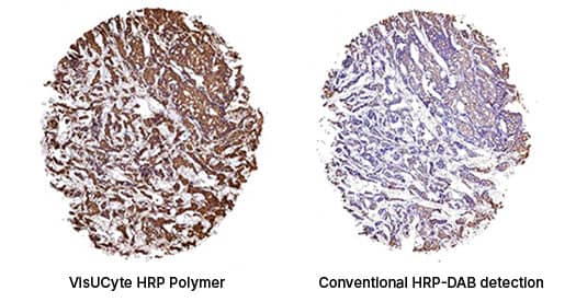
Figure 2. Side-by-side comparison of staining using different detection chemistries on adjacent tissue sections of human breast cancer. Tissue sections were first incubated with primary antibodies and then subjected to staining development using either a Goat VisUCyte kit (Catalog# VCTS004B) or conventional HRP-DAB detection chemistry. Strong specific tissue staining is clearly seen using the VisUCyte kit whereas conventional HRP-DAB detection produced little or no staining. In clinical research using VisUCyte kits allows for high-sensitivity detection of cancer biomarkers compared to conventional HRP-DAB protocol.
3. Streamlined Protocol: The VisUCyte™ kits offer a time-efficient staining procedure, completing IHC experiments in as little as 1-2 hours without compromising the quality of results. View detailed tissue staining protocols.
4. Ready-to-Use Convenience: The kits come with pre-optimized reagents, eliminating the need for intricate staining optimization, and providing researchers with a hassle-free experience.
5. Versatility: VisUCyte™ kits are compatible with a wide range of tissue types, making them suitable for various research applications, from basic research to clinical diagnostics.
A Leap Forward in Tissue Staining
Bio-Techne's VisUCyte™ HRP Polymer Conjugated Secondary Antibodies represent a remarkable advancement in the field of IHC. With its high sensitivity, lack of non-specific staining, and efficient protocol, these kits streamline the process of detecting protein targets in tissue sections. Researchers can now enjoy improved accuracy, reduced experimental times, and cost-effective staining, all while gaining deeper insights into the intricate world of tissue-specific protein expression.

Product Marketing Specialist at Bio-Techne