Human Aminopeptidase N/CD13 Antibody
R&D Systems, part of Bio-Techne | Catalog # AF3815


Key Product Details
Validated by
Species Reactivity
Validated:
Cited:
Applications
Validated:
Cited:
Label
Antibody Source
Product Specifications
Immunogen
Lys69-Lys967
Accession # P15144
Specificity
Clonality
Host
Isotype
Scientific Data Images for Human Aminopeptidase N/CD13 Antibody
Detection of Human Aminopeptidase N/CD13 by Western Blot.
Western blot shows lysates of human kidney tissue and human prostate tissue. PVDF membrane was probed with 0.5 µg/mL of Sheep Anti-Human Aminopeptidase N/CD13 Antigen Affinity-purified Polyclonal Antibody (Catalog # AF3815) followed by HRP-conjugated Anti-Sheep IgG Secondary Antibody (Catalog # HAF016). A specific band was detected for Aminopeptidase N/CD13 at approximately 150 kDa (as indicated). This experiment was conducted under reducing conditions and using Immunoblot Buffer Group 1.Detection of Aminopeptidase N/CD13 in U937 Human Cell Line by Flow Cytometry.
U937 human histiocytic lymphoma cell line was stained with Sheep Anti-Human Aminopeptidase N/CD13 Antigen Affinity-purified Polyclonal Antibody (Catalog # AF3815, filled histogram) or isotype control antibody (Catalog # 5-001-A, open histogram), followed by APC-conjugated Anti-Sheep IgG Secondary Antibody (Catalog # F0127). View our protocol for Staining Membrane-associated Proteins.Aminopeptidase N/CD13 in Human Kidney.
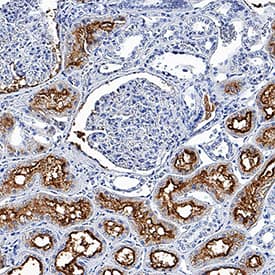
Aminopeptidase N/CD13 was detected in immersion fixed paraffin-embedded sections of human kidney using Sheep Anti-Human Aminopeptidase N/CD13 Antigen Affinity-purified Polyclonal Antibody (Catalog # AF3815) at 0.3 µg/mL overnight at 4 °C. Tissue was stained using the Anti-Sheep HRP-DAB Cell & Tissue Staining Kit (brown; Catalog # CTS019) and counterstained with hematoxylin (blue). Specific staining was localized to convoluted tubules. View our protocol for Chromogenic IHC Staining of Paraffin-embedded Tissue Sections.Applications for Human Aminopeptidase N/CD13 Antibody
CyTOF-ready
Flow Cytometry
Sample: U937 human histiocytic lymphoma cell line
Immunohistochemistry
Sample: Immersion fixed paraffin-embedded sections of human kidney
Immunoprecipitation
Sample: Conditioned cell culture medium spiked with Recombinant Human Aminopeptidase N/CD13 (Catalog # 3815-ZN), see our available Western blot detection antibodies
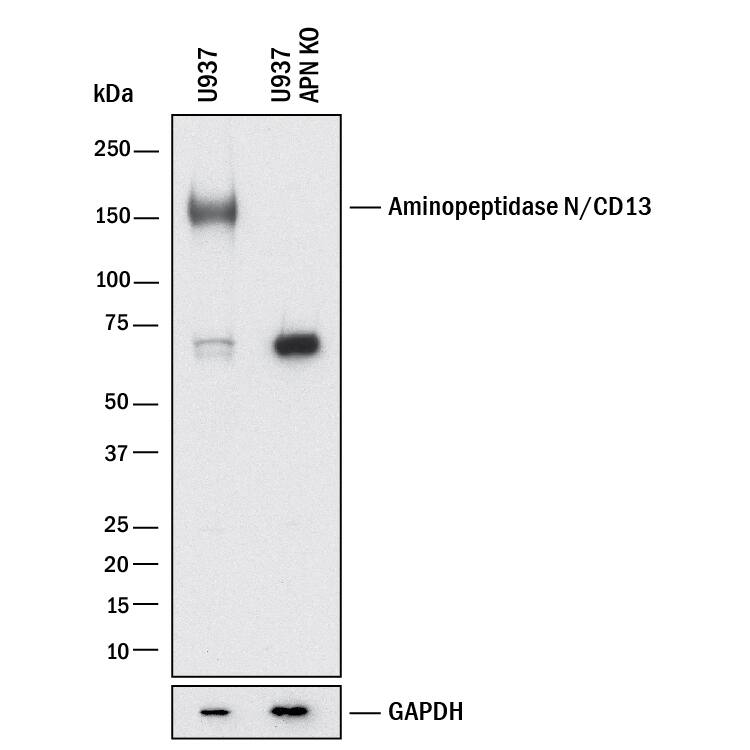
Knockout Validated
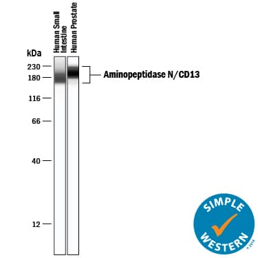
Simple Western
Sample: Human small intestine tissue and Human prostate tissue
Western Blot
Sample: Human kidney tissue and human prostate tissue
Formulation, Preparation, and Storage
Purification
Reconstitution
Formulation
Shipping
Stability & Storage
- 12 months from date of receipt, -20 to -70 °C as supplied.
- 1 month, 2 to 8 °C under sterile conditions after reconstitution.
- 6 months, -20 to -70 °C under sterile conditions after reconstitution.
Background: Aminopeptidase N/CD13
The human ANPEP gene encodes aminopeptidase N (APN), which is also known as microsomal aminopeptiase, alanyl aminopeptidase, aminopeptidase M, CD13, or membrane protein p161 (1‑3). The deduced amino acid sequence of human APN consists of a short cytoplasmic tail (residues 2 to 8), a transmembrane region (residue 9 to 32), a Ser/Thr rich region and a zinc metalloprotease domain (residues 69 to 966). Widely expressed in many cells, tissues and species, APN cleaves the N-terminal amino acids from bioactive peptides, leading to their inactivation or degradation. The roles of APN in many fields, such as neuroscience, hematopoeitic cells, immune system, angiogenesis, cancer and viral infection, have been reviewed (3).
References
- Olsen, J. et al. (1988) FEBS Lett. 238:307.
- Look, A.T. et al. (1989) J. Clin. Invest. 83:1299.
- Turner, A.J. (2004) in Handbook of Proteolytic Enzymes (ed. Barrett, et al.) pp. 289, Academic Press, San Diego.
Alternate Names
Gene Symbol
UniProt
Additional Aminopeptidase N/CD13 Products
Product Documents for Human Aminopeptidase N/CD13 Antibody
Product Specific Notices for Human Aminopeptidase N/CD13 Antibody
For research use only


