Human Granzyme B Antibody
R&D Systems, part of Bio-Techne | Catalog # MAB2906


Key Product Details
Species Reactivity
Validated:
Cited:
Applications
Validated:
Cited:
Label
Antibody Source
Product Specifications
Immunogen
Gly19-Tyr247
Accession # P10144
Specificity
Clonality
Host
Isotype
Scientific Data Images for Human Granzyme B Antibody
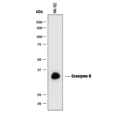
Detection of Human Granzyme B by Western Blot.
Western blot shows lysate of NK-92 human natural killer lymphoma cell line. PVDF membrane was probed with 0.5 µg/mL of Mouse Anti-Human Granzyme B Monoclonal Antibody (Catalog # MAB2906) followed by HRP-conjugated Anti-Mouse IgG Secondary Antibody (Catalog # HAF018). A specific band was detected for Granzyme B at approximately 32-35 kDa (as indicated). This experiment was conducted under reducing conditions and using Immunoblot Buffer Group 1.Granzyme B in Human PBMCs.
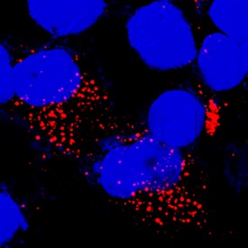
Granzyme B was detected in immersion fixed human peripheral blood mononuclear cells (PBMCs) using Mouse Anti-Human Granzyme B Monoclonal Antibody (Catalog # MAB2906) at 8 µg/mL for 3 hours at room temperature. Cells were stained using the NorthernLights™ 557-conjugated Anti-Mouse IgG Secondary Antibody (red; Catalog # NL007) and counterstained with DAPI (blue). Specific staining was localized to cytoplasm. View our protocol for Fluorescent ICC Staining of Non-adherent Cells.Detection of Human Granzyme B by Simple WesternTM.
Simple Western lane view shows lysates of NK-92 human natural killer lymphoma cell line, loaded at 0.2 mg/mL. A specific band was detected for Granzyme B at approximately 42 kDa (as indicated) using 5 µg/mL of Mouse Anti-Human Granzyme B Monoclonal Antibody (Catalog # MAB2906) . This experiment was conducted under reducing conditions and using the 12-230 kDa separation system.Applications for Human Granzyme B Antibody
CyTOF-ready
Immunocytochemistry
Sample: Immersion fixed human peripheral blood mononuclear cells
Immunohistochemistry
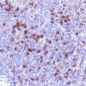
Sample: Immersion fixed paraffin-embedded sections of human tonsil
Intracellular Staining by Flow Cytometry
Sample: NK-92 human natural killer lymphoma cell line fixed with paraformaldehyde and permeabilized with saponin
Simple Western
Sample: NK‑92 human natural killer lymphoma cell line
Western Blot
Sample: NK‑92 human natural killer lymphoma cell line
Formulation, Preparation, and Storage
Purification
Reconstitution
Formulation
Shipping
Stability & Storage
- 12 months from date of receipt, -20 to -70 °C as supplied.
- 1 month, 2 to 8 °C under sterile conditions after reconstitution.
- 6 months, -20 to -70 °C under sterile conditions after reconstitution.
Background: Granzyme B
Granzyme B is a member of the granzyme family of the serine proteases found specifically in the cytotoxic granules of cytotoxic T lymphocytes (CTL) and natural killer (NK) cells (1, 2). Granzyme B plays an essential role in granule-mediated apoptosis and may have additional roles in rheumatoid arthritis and in bacterial and viral infections (3). It activates various caspases and cleaves proteins such as aggrecan (3). Human Granzyme B is synthesized as a precursor (247 residues) with a signal peptide (residues 1-18), a pro peptide (residues 19-20), and a mature chain (residues 21-247) (4-6). The recombinant human (rh) Granzyme B consisting of residues 19-247 was expressed and purified. After being activated by active cathepsin C, rhGranzyme B cleaves a thioester substrate described previously (3).
References
- Kam, C-M. et al. (2000) Biochim. Biophys. Acta 1477:307.
- Smyth, M.J. et al. (1996) J. Leukoc. Biol. 60:555.
- Froelich, C.J. (2004) in Handbook of Proteolytic Enzymes, Barrett, A.J. et al. eds. pp. 1549.
- Schmid, J. and C. Weissman (1987) J. Immunol. 139:250.
- Caputo, A. et al. (1988) J. Biol. Chem. 263:6363.
- Trapani, J.A. et al. (1988) Proc. Natl. Acad. Sci. USA 85:6924.
Alternate Names
Gene Symbol
UniProt
Additional Granzyme B Products
Product Documents for Human Granzyme B Antibody
Product Specific Notices for Human Granzyme B Antibody
For research use only


