Human/Mouse/Rat RelA/NF kappaB p65 Antibody
R&D Systems, part of Bio-Techne | Catalog # MAB5078

Key Product Details
Validated by
Species Reactivity
Validated:
Cited:
Applications
Validated:
Cited:
Label
Antibody Source
Product Specifications
Immunogen
Asn456-Ser551
Accession # Q04206
Specificity
Clonality
Host
Isotype
Scientific Data Images for Human/Mouse/Rat RelA/NF kappaB p65 Antibody
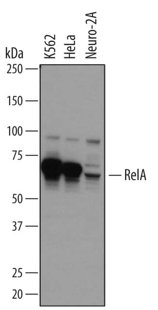
Detection of Human and Mouse RelA/NF kappaB p65 by Western Blot.
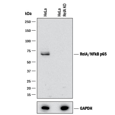
Western blot shows lysates of K562 human chronic myelogenous leukemia cell line, HeLa human cervical epithelial carcinoma cell line, and Neuro-2A mouse neuroblastoma cell line. PVDF membrane was probed with 2 µg/mL of Mouse Anti-Human/Mouse/Rat RelA/NF kappa B p65 Monoclonal Antibody (Catalog # MAB5078) followed by HRP-conjugated Anti-Mouse IgG Secondary Antibody (Catalog # HAF007). A specific band was detected for RelA/NF kappa B p65 at approximately 70 kDa (as indicated). This experiment was conducted under reducing conditions and using Immunoblot Buffer Group 1.Detection of Rat RelA/NF kappaB p65 by Western Blot.
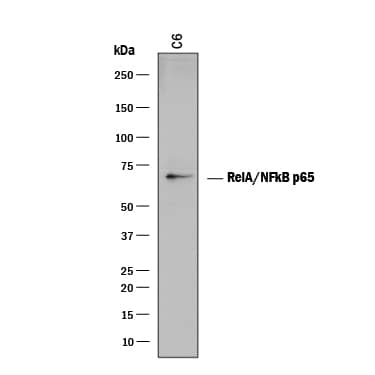
Western blot shows lysates of C6 rat glioma cell line. PVDF membrane was probed with 2 µg/mL of Mouse Anti-Human/Mouse/Rat RelA/NF kappa B p65 Monoclonal Antibody (Catalog # MAB5078) followed by HRP-conjugated Anti-Mouse IgG Secondary Antibody (Catalog # HAF018). A specific band was detected for RelA/NF kappa B p65 at approximately 65 kDa (as indicated). This experiment was conducted under reducing conditions and using Immunoblot Buffer Group 1.RelA/NF kappaB p65 in K562 Human Cell Line.
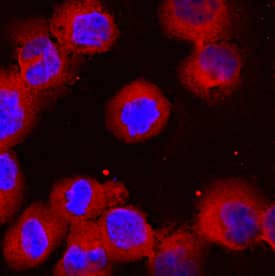
RelA/NF kappa B p65 was detected in immersion fixed K562 human chronic myelogenous leukemia cell line using Mouse Anti-Human/Mouse/Rat RelA/NF kappa B p65 Monoclonal Antibody (Catalog # MAB5078) at 10 µg/mL for 3 hours at room temperature. Cells were stained using the NorthernLights™ 557-conjugated Anti-Mouse IgG Secondary Antibody (red; Catalog # NL007) and counterstained with DAPI (blue). View our protocol for Fluorescent ICC Staining of Non-adherent Cells.Applications for Human/Mouse/Rat RelA/NF kappaB p65 Antibody
CyTOF-ready
Immunocytochemistry
Sample: Immersion fixed K562 human chronic myelogenous leukemia cell line
Intracellular Staining by Flow Cytometry
Sample: HeLa human cervical epithelial carcinoma cell line, fixed with paraformaldehyde and permeabilized with methanol
Knockout Validated
Western Blot
Sample: K562 human chronic myelogenous leukemia cell line, HeLa human cervical epithelial carcinoma cell line, Neuro‑2A mouse neuroblastoma cell line, and C6 rat glioma cell line
Reviewed Applications
Read 2 reviews rated 5 using MAB5078 in the following applications:
Formulation, Preparation, and Storage
Purification
Reconstitution
Formulation
Shipping
Stability & Storage
- 12 months from date of receipt, -20 to -70 °C as supplied.
- 1 month, 2 to 8 °C under sterile conditions after reconstitution.
- 6 months, -20 to -70 °C under sterile conditions after reconstitution.
Background: RelA/NFkB p65
RelA p65 (v-rel reticuloendotheliosis viral oncogene homolog A) is a 65 kDa member of the NF kappaB family of nuclear transcription factors. Dimers of p65 with the p50 subunit are the most common form of the NF kappaB transcription factor, but dimers with it or other family members can also occur. Upon activation, RelA p65 forms an heterotetramer and moves into the nucleus where it binds to specific DNA sequences. An alternatively spliced isoform that lacks amino acids (aa) 222‑231 (p65 delta) does not bind DNA. Over the sequence used as an immunogen, human RelA p65 shares 96% and 98% aa identity with mouse and rat RelA p65, respectively. This portion includes one of eight potential ser/thr phosphorylation sites, two acetylation sites, and most of the Rel homology domain that interacts with I kappaB inhibitors.
Long Name
Alternate Names
Entrez Gene IDs
Gene Symbol
UniProt
Additional RelA/NFkB p65 Products
Product Documents for Human/Mouse/Rat RelA/NF kappaB p65 Antibody
Product Specific Notices for Human/Mouse/Rat RelA/NF kappaB p65 Antibody
For research use only
