Human TM4SF1/L6 Antibody
R&D Systems, part of Bio-Techne | Catalog # MAB8164

Key Product Details
Species Reactivity
Validated:
Cited:
Applications
Validated:
Cited:
Label
Antibody Source
Product Specifications
Immunogen
Met1-Cys202
Accession # P30408
Specificity
Clonality
Host
Isotype
Scientific Data Images for Human TM4SF1/L6 Antibody
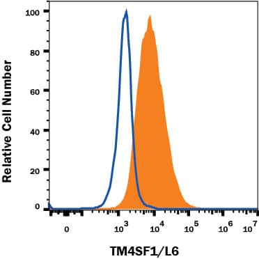
Detection of TM4SF1/L6 in A549 Human Cell Line by Flow Cytometry.
A549 human lung carcinoma cell line was stained with Mouse Anti-Human TM4SF1/L6 Monoclonal Antibody (Catalog # MAB8164, filled histogram) or isotype control antibody (Catalog # MAB002, open histogram), followed by Allophycocyanin-conjugated Anti-Mouse IgG Secondary Antibody (Catalog # F0101B). View our protocol for Staining Membrane-associated Proteins.Detection of TM4SF1/L6 in HEK293 Human Cell Line Transfected with Human TM4SF1/L6 and eGFP by Flow Cytometry.
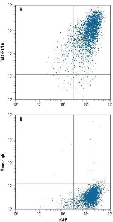
HEK293 human embryonic kidney cell line transfected with human TM4SF1/L6 and eGFP was stained with either (A) Mouse Anti-Human TM4SF1/L6 Monoclonal Antibody (Catalog # MAB8164) or (B) Mouse IgG1Isotype Control (Catalog # MAB002) followed by Phycoerythrin-conjugated Anti-Mouse IgG Secondary Antibody (Catalog # F0102B). View our protocol for Staining Membrane-associated Proteins.TM4SF1/L6 in A549 Human Cell Line.
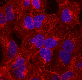
TM4SF1/L6 was detected in immersion fixed A549 human lung carcinoma cell line using Mouse Anti-Human TM4SF1/L6 Monoclonal Antibody (Catalog # MAB8164) at 10 µg/mL for 3 hours at room temperature. Cells were stained using the NorthernLights™ 557-conjugated Anti-Mouse IgG Secondary Antibody (red; Catalog # NL007) and counterstained with DAPI (blue). Specific staining was localized to cell membranes. View our protocol for Fluorescent ICC Staining of Cells on Coverslips.Applications for Human TM4SF1/L6 Antibody
CyTOF-ready
Flow Cytometry
Sample: A549 human lung carcinoma cell line and HEK293 human embryonic kidney cell line transfected with human TM4SF1/L6 and eGFP
Immunocytochemistry
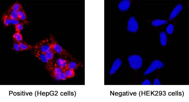
Sample: Immersion fixed A549 human lung carcinoma cell line and immersion fixed HepG2 human hepatocellular carcinoma cell line
Immunohistochemistry
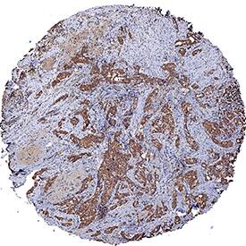
Sample: Immersion fixed paraffin-embedded sections of human breast cancer tissue
Reviewed Applications
Read 1 review rated 5 using MAB8164 in the following applications:
Formulation, Preparation, and Storage
Purification
Reconstitution
Formulation
Shipping
Stability & Storage
- 12 months from date of receipt, -20 to -70 °C as supplied.
- 1 month, 2 to 8 °C under sterile conditions after reconstitution.
- 6 months, -20 to -70 °C under sterile conditions after reconstitution.
Background: TM4SF1/L6
|
TM4SF1 (Transmembrane 4 L6 family Member 1; also L6 and M3s1) is a 23-28 kDa member of the L6 tetraspanin family of molecules. It is expressed by fibroblasts, endothelial cells and a variety of tumor cells. TM4SF1 is embedded in both the plasma membrane and the membrane of late endocytic organelles. Here, it appears to be ubiquitinated, and to regulate endocytosis, an action that impacts effective cell migration. TM4SF1 is reported to interact with alpha5 and beta1 integrins, and this may impact cell motility. It also suppresses the expression of CD63 and CD82, two molecules that impede cell mobility. Mouse TM4SF1 is a 202 amino acid (aa) 4-transmembrane (TM) glycoprotein. It contains a short N-terminal cytoplasmic region (aa 1-9), followed by four TM regions and another C-terminal cytoplasmic region (aa 183-202). There is one potential isoform variant that shows an alternative start site at Met60. Over aa 116-161, mouse TM4SF1 shares 73% and 96% aa sequence identity with human and rat TM4SF1, respectively. |
Long Name
Alternate Names
Gene Symbol
UniProt
Additional TM4SF1/L6 Products
Product Documents for Human TM4SF1/L6 Antibody
Product Specific Notices for Human TM4SF1/L6 Antibody
For research use only
