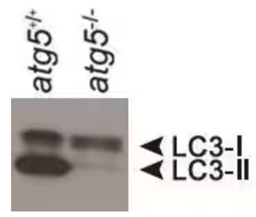
Microtubule-associated protein light chain 3 (LC3) is considered one of the definitive markers of autophagy, and its use is widespread in labs throughout the world. Despite its popularity, there are several considerations when employing LC3 antibodies in immunoassays, Western blots in particular.
LC3 is expressed as a propeptide and is subsequently cleaved to form LC3-I. Initiation of autophagy causes the conversion of LC3-I to LC3-II via the addition of a phosphatidylethanolamine (PE) group to the C terminus. The PE group increases the rate of band migration in an SDS-PAGE gel, likely due to its hydrophobic nature; this modification commonly manifests as the appearance of a doublet in a Western blot. The lipophilic character of the PE group also facilitates the insertion of LC3-II into the membranes of autophagosomes, and as a result, LC3-II is degraded as autophagosomes are turned over. An increase in LC3-II band intensity and a decrease in LC3-I expression is considered the hallmark of autophagy but increases in LC3-II can be caused by enhanced autophagosome synthesis or reduced autophagosome recycling, making interpreting LC3 staining misleading.
Analysis of LC3 Western blots is further complicated by a difference in the affinities of antibodies for LC3-I and LC3-II and by the marked difference in LC3 expression in varying cell types and tissues. Due to these factors, a consensus has emerged that the total amount of LC3-II should only be evaluated and compared to a loading control as a means of assessing autophagy. In addition, many investigators include control extracts collected from cells treated with various inhibitors in order to determine the efficiency of autophagic flux in their experimental samples. The LC3 expression pattern in these controls ideally indicates the cause of LC3-II accumulation (or its absence) depending on the type of inhibitor used. One last consideration is the measurement of autophagic flux over a period of time as opposed to harvesting extracts from one static point. For example, although the shift from LC3-I to LC3-II is evident following short periods of stress, the LC3 signal disappears after extended starvation in many cell types.
For LC3 troubleshooting tips and scientific technical answers to 25+ frequently asked questions (FAQs) on autophagy research, read our FAQs - Autophagy and LC3.
Bio-Techne offers LC3 reagents for your research needs including:
