Human/Mouse IGF-I R/IGF1R Antibody
R&D Systems, part of Bio-Techne | Catalog # MAB391


Key Product Details
Species Reactivity
Validated:
Cited:
Applications
Validated:
Cited:
Label
Antibody Source
Product Specifications
Immunogen
Glu31-Asn932
Accession # P08069
Specificity
Clonality
Host
Isotype
Endotoxin Level
Scientific Data Images for Human/Mouse IGF-I R/IGF1R Antibody
Detection of Human IGF-I R/IGF1R by Western Blot.
Western blot shows lysates of NTera-2 human testicular embryonic carcinoma cell line, SK-Mel-28 human malignant melanoma cell line, and G361 human melanoma cell line. PVDF membrane was probed with 1 µg/mL of Mouse Anti-Human/Mouse IGF-I R/IGF1R Monoclonal Antibody (Catalog # MAB391) followed by HRP-conjugated Anti-Mouse IgG Secondary Antibody (Catalog # HAF007). A specific band was detected for IGF-I R/IGF1R at approximately 275 kDa (as indicated). This experiment was conducted under non-reducing conditions and using Immunoblot Buffer Group 2.Detection of IGF-I R/IGF1R in MCF‑7 Human Cell Line by Flow Cytometry.
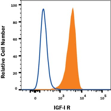
MCF-7 human breast cancer cell line was stained with Mouse Anti-Human IGF-I R/IGF1R Monoclonal Antibody (Catalog # MAB391, filled histogram) or isotype control antibody (Catalog # MAB002, open histogram) followed by anti-mouse IgG PE-conjugated secondary antibody (Catalog # F0102B). View our protocol for Staining Membrane-associated Proteins.IGF-I R/IGF1R in MCF‑7 and HDLM Human Cell Lines.
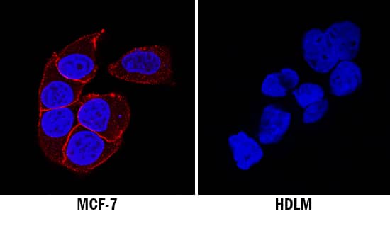
IGF-I R/IGF1R was detected in immersion fixed MCF-7 human breast cancer cell line (positive staining; left panel) and HDLM human Hodgkin's lymphoma cell line (negative staining; right panel) using Mouse Anti-Human/Mouse IGF-I R/IGF1R Monoclonal Antibody (Catalog # MAB391) at 3 µg/mL for 3 hours at room temperature. Cells were stained using the NorthernLights™ 557-conjugated Anti-Mouse IgG Secondary Antibody (red; Catalog # NL007) and counterstained with DAPI (blue). Specific staining was localized to cell membrane in MCF-7 cell line. View our protocol for Fluorescent ICC Staining of Cells on Coverslips.Applications for Human/Mouse IGF-I R/IGF1R Antibody
Flow Cytometry
Sample: MCF-7 human breast cancer cell line
Immunocytochemistry
Sample: Immersion fixed MCF‑7 human breast cancer cell line
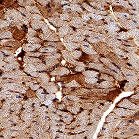
Immunohistochemistry
Sample: Perfusion fixed paraffin-embedded sections of mouse heart
Western Blot
Sample: NTera-2 human testicular embryonic carcinoma cell line, SK-Mel-28 human malignant melanoma cell line, and G361 human melanoma cell line under non-reducing conditions only
Neutralization
Human IGF-I R/IGF1R Sandwich Immunoassay
Reviewed Applications
Read 1 review rated 5 using MAB391 in the following applications:
Formulation, Preparation, and Storage
Purification
Reconstitution
Formulation
Shipping
Stability & Storage
- 12 months from date of receipt, -20 to -70 °C as supplied.
- 1 month, 2 to 8 °C under sterile conditions after reconstitution.
- 6 months, -20 to -70 °C under sterile conditions after reconstitution.
Background: IGF-I R/IGF1R
Insulin-like growth factor I receptor (IGF-I R) is a disulfide-linked heterotetrameric transmembrane protein consisting of two alpha and two beta subunits. Both the alpha and beta subunits are encoded within a single receptor precursor cDNA. The proreceptor polypeptide is proteolytically cleaved and disulfide-linked to yield the mature heterotetrameric receptor. The alpha subunit of IGF-I R is extracellular while the beta subunit has an extracellular domain, a transmembrane domain and a cytoplasmic tyrosine kinase domain. IGF-I R is highly expressed in all cell types and tissues. Essentially all of the biological activities of IGF-I and -II have been shown to be mediated via IGF-I R.
Long Name
Alternate Names
Entrez Gene IDs
Gene Symbol
UniProt
Additional IGF-I R/IGF1R Products
Product Documents for Human/Mouse IGF-I R/IGF1R Antibody
Product Specific Notices for Human/Mouse IGF-I R/IGF1R Antibody
For research use only


